Using a Thermal Image camera to enhance the management of the at-risk limb in a remote and rural region
Treating diabetic foot problems in patients in remote and rural areas
is challenging.(1) In NHS Highland, we tested the use of digital thermography in the high-risk foot clinic, as a diagnostic tool to assist in the early diagnosis of foot complications including sub-clinical infections and diabetic foot ulcers; and in patients with suspected osteomyelitis or Charcot.
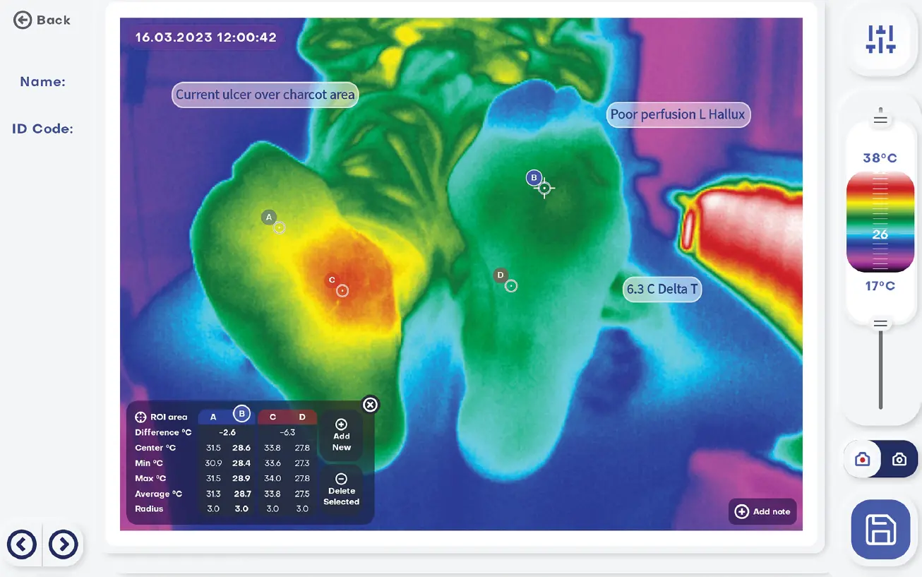
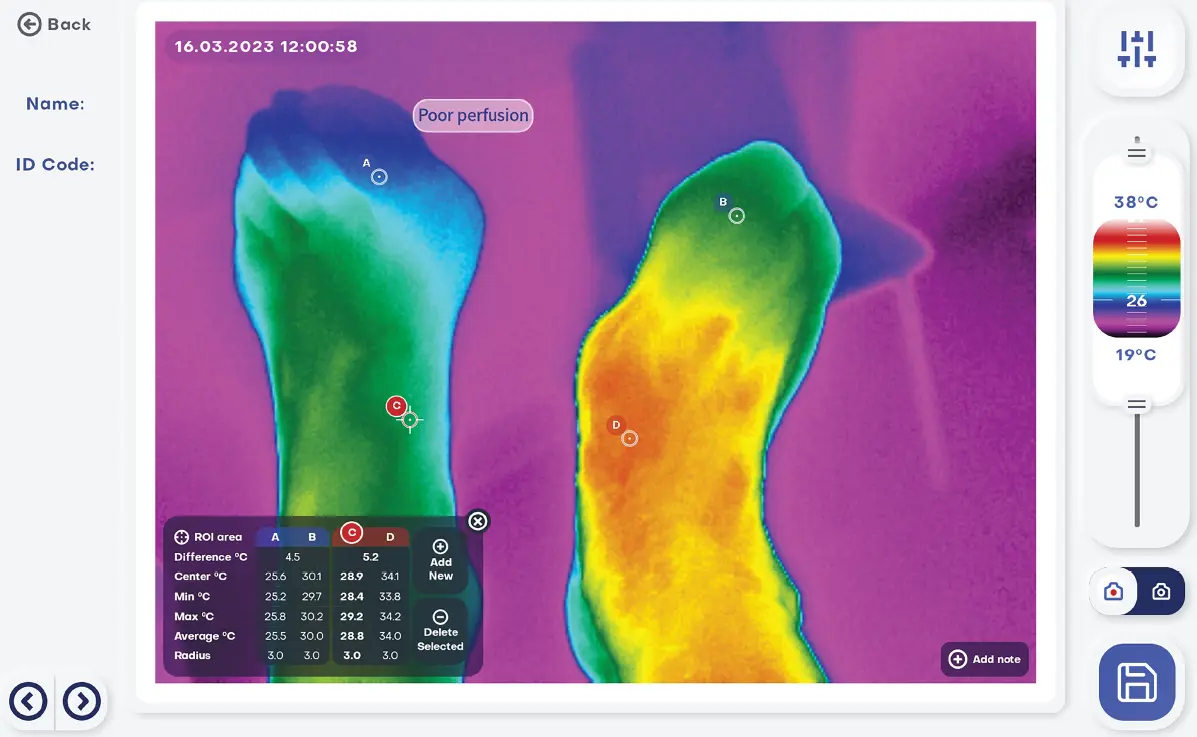
A thermal imaging camera is designed to enhance clinical practice in the management of the at-risk limb. Detecting increased temperature regions in tissue before any visible inflammatory signs may assist in reducing the development of more complex medical conditions.
The use of thermal imaging can also aid in the identification of areas of stress with the potential for tissue breakdown, to indicate ‘hot spots’ which require off-loading or orthotic modifications. The presence of cold spots can also indicate peripheral arterial disease.
Method
An Android tablet with a thermal camera was used in the high-risk foot clinic. Patients were scanned with the thermal camera to detect thermal anomalies between the limbs, with special interest paid to elevated temperature asymmetries more than 2.2 °C which has been reported to be indicative of potential infections and an early sign of incipient diabetes foot ulcers.
The camera was also used during a video consultation.
Results
By scanning patients in the clinic and in their homes, we were able to make faster medical decisions, resulting in better outcomes, decreasing the use of expensive diagnostic exams, and in one case avoid a toe amputation. Furthermore, the multi-disciplinary team acted quickly to ensure the correct antibiotic therapy was in place to effectively manage a potential deep bone infection.
The intuitive thermal images motivated the patients in behavioural change, adapting their lifestyle, and reinforced better collaboration with the foot team working towards wound healing.
Discussion
Thermal imaging enabled enhanced clinical decision making, and can be a great patient education tool encouraging behavioural change.
The use of thermography has the potential to enhance the quality of the established RAPID pathway(1) for remote monitoring and management of patients with active foot wounds in a geographically challenged region.
This revolutionary diagnostic tool has the potential to give podiatrists confidence in early diagnosis and identifying and managing foot infections more effectively. We appreciate it is a small study but shows great potential and so would benefit from further investigation in a larger and more diverse population.