
Lower-Limb Thermal Imaging

Thermidas thermal imaging provides a fast, non‑invasive way to detect inflammation, vascular changes, and early tissue stress in the lower limbs. It helps clinicians identify complications, such as diabetic foot issues, before symptoms appear, supporting earlier intervention and improved patient outcomes.

Clinical Challenges in Lower-Limb Care

  1. Diabetic Foot Complications
    Loss of protective sensation and impaired wound healing make early detection essential.
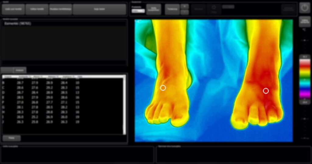
    Studies show that localized temperature increases can precede ulceration by 1–5 weeks in high‑risk patients (Armstrong et al., 2007; Lavery et al., 2004). Contralateral temperature differences ≥ 2.2°C are associated with elevated ulcer risk.
  2. Vascular Disorders
    Peripheral arterial disease (PAD) and microvascular insufficiency can present as cooler regions due to reduced perfusion.
    Thermography may support clinical evaluation by visualizing temperature asymmetry linked to circulatory compromise.
    (Frick et al., 2022).
  3. Musculoskeletal and Overuse Injuries
    Acute or chronic loading of the foot and ankle often produces characteristic heat patterns.
    Research indicates that thermographic asymmetries correlate with tissue stress and inflammatory response.
    (Sillero‑Quintana et al., 2017).

Infrared thermology is a powerful detector of problems that affect a patient’s physiology.

Thermidas provides revolutionary, non-contact, non-invasive, physiological diagnostic tools utilizing thermography to identify and diagnose underlying pathology quickly and reliably at earlier stages than currently adopted methods.

Jones BF. A reappraisal of the use of infrared thermal image analysis in medicine. IEEE Trans Med Imaging. 1998 Dec;17(6):1019-27. doi: 10.1109/42.746635. PMID: 10048859.

Case examples of thermographic use​

Lower-limb complications are common in patients with diabetes, vascular insufficiency, neuropathy, and musculoskeletal injuries. Many of these conditions begin with subtle physiological changes—especially localized temperature variation—well before visible symptoms develop.

Thermidas thermal imaging provides clinicians with standardized plantar and dorsal views of the feet and lower legs, enabling early detection of abnormal temperature patterns that may indicate inflammation, impaired perfusion, or tissue stress.

Case example

Broken / operated Achilles tendon

  • First thermal image taken eight hours after Orthosis was removed
  • Second image taken six weeks later

Case example

Stress fracture

  • The patient felt pain on the left foot
  • Thermal imaging revealed a substantial asymmerty in the temperature
  • Later a stress fracture was diagnosed

Case example

Insoles' effectiveness

  • Patient is complaining about pain in the knee area
  • Thermal imaging revealed abnormal temperatures and helped locate the problem areas
  • Custom-made insoles were made
  • Follow-up thermal images were taken to evaluate the effectiveness of the insoles
  • The situation had calmed down, and the patient was able to perform normal daily activities

Recommended solution

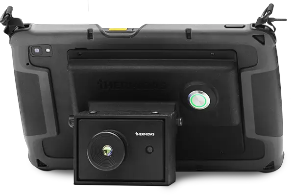
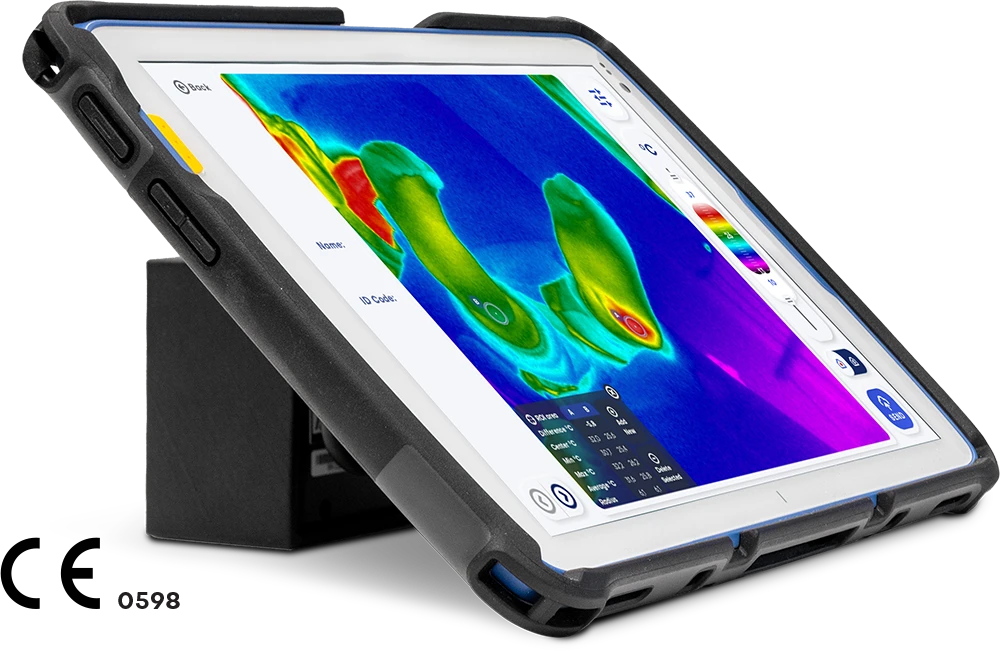
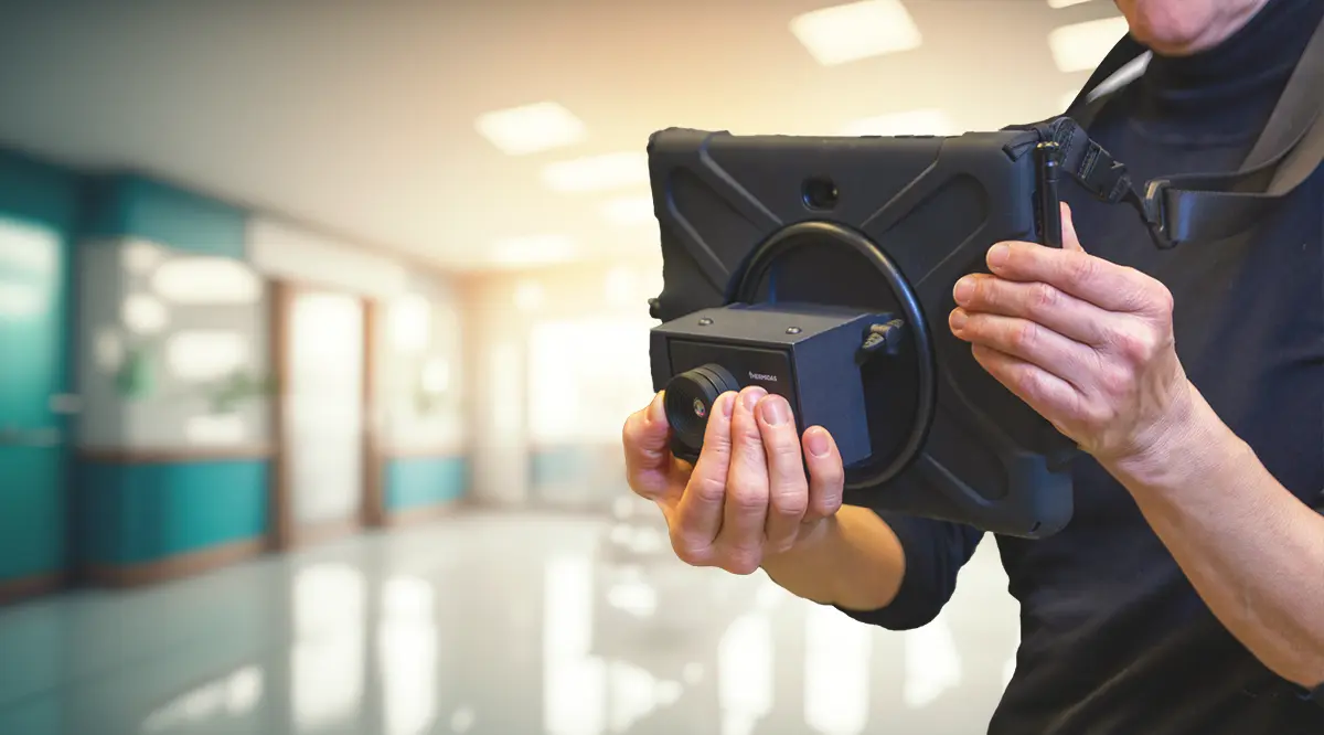
Thermidas IRT-384 Tablet

The IRT-384 Tablet is a high-quality thermal imaging solution for locating problem areas and monitoring the effectiveness of treatment.

Thermal imaging also supports the client’s understanding of rehabilitation progress by providing clear, objective information on the effects of treatment.

Case example

CRPS

  • The patient had a toy dropped on her left foot​
  • Nothing was detected in the previous X-ray or MRI sessions​
  • In the thermal imaging, a temperature difference of over 3 degrees was detected ​
  • CRPS on the left foot was later diagnosed​

How can we help you?