
Clinical Problem
Skin irritation, inflammation, and early tissue breakdown can develop inside prosthetic sockets and orthotic devices long before visible signs appear. These issues are frequently caused by:
- Localized mechanical loading
- Poor or changing socket fit
- Friction or shear between skin, liner, and device interface
- Reduced protective sensation (e.g., diabetic patients)
- Changes in activity, weight, or limb volume
Such problems can lead to discomfort, recurrent clinic visits, reduced device adherence, and progression toward more serious skin injury.
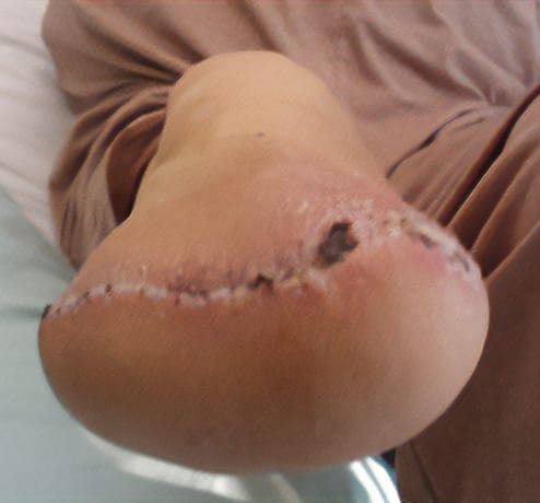
Although clinicians routinely rely on patient history, palpation, and visual inspection, these methods may not reveal early inflammatory changes or loading patterns hidden beneath the device.
What thermal patterns may indicate?
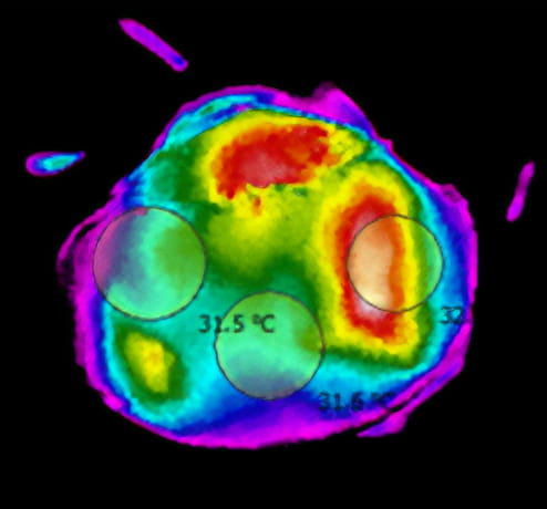
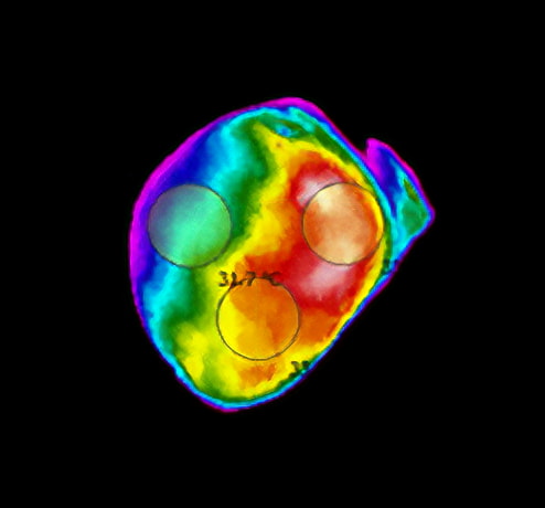
Thermidas medical-grade thermal imaging provides an additional, non‑contact method for visualizing temperature patterns associated with potential mechanical stress or inflammation.
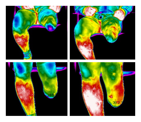
- Temperature asymmetry may be associated with increased loading or early inflammatory response.
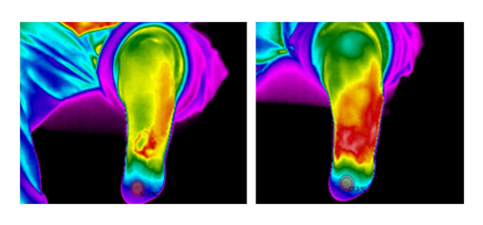
- Localized hotspots may correspond to areas of friction or pressure.
- Cool areas can reflect reduced perfusion or off‑loading.
- Changes across visits may provide insight into socket fit issues, healing, or treatment respons
These patterns are intended to support, not replace, clinical judgment. They serve as objective adjunctive information that can complement examination, patient‑reported symptoms, and device evaluation.
Prosthetics
Thermal imaging may support clinicians in evaluating:
- Residual limb temperature distribution before and after activity
- Areas potentially subject to excessive socket pressure
- Fit issues related to limb volume changes
- Monitoring of high‑risk patients with reduced sensation
- Post‑adjustment outcomes and follow‑up documentation
Orthotics
Thermal imaging may help visualize:
- Mechanical loading patterns in orthoses
- Skin stress zones under foot orthoses, AFOs, spinal orthoses, or pressure‑relief devices
- Evolution of inflammatory patterns over time
- Response to modifications in trim lines, padding, or materials
Recommended solution
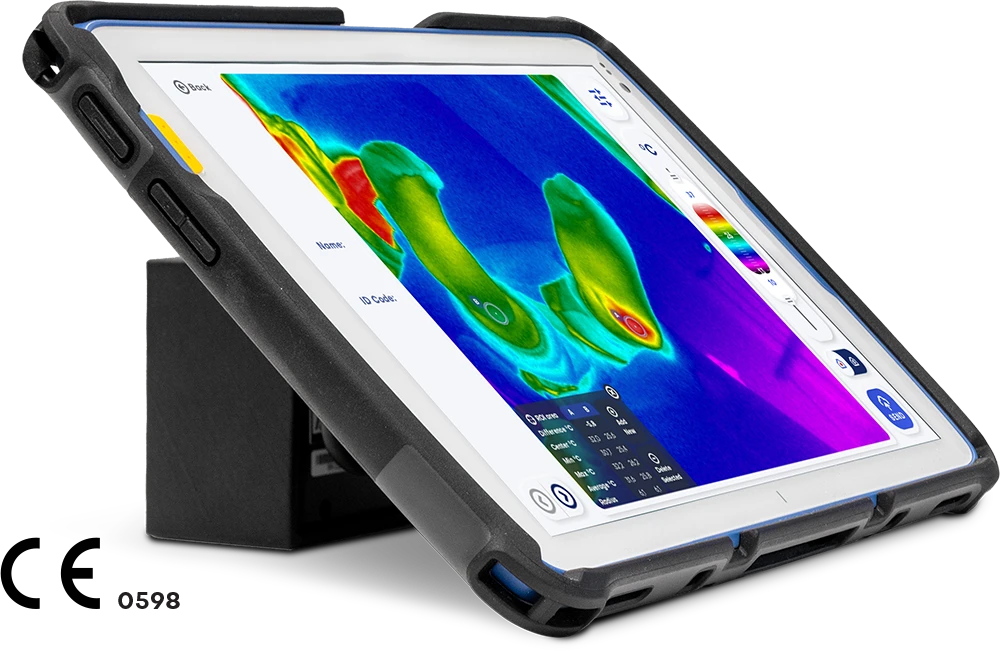
Thermidas IRT-384 Tablet
The IRT-384 Tablet is a high-quality thermal imaging solution for locating problem areas and monitoring the effectiveness of treatment.
Thermal imaging also supports the client’s understanding of rehabilitation progress by providing clear, objective information on the effects of treatment.
The benefits of thermal imaging for prosthetics
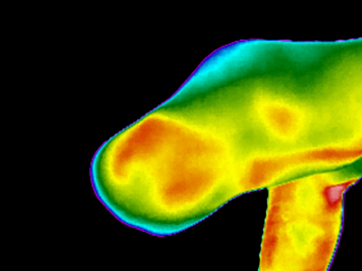
The quantitative measure of the state of the residual limb perfusion which is revealed by thermal maps may provide important information about:
- Existing defects of the socket, that during walking translate into excessive forces and in turn temperature increase
- Skin inflammations, e.g., as the result of the liner leading to excessive humidity and heating
- The classification and treatment of phantom limb pain
Clinical workflow example
Thermidas imaging can be integrated into routine prosthetics & orthotics consultations with minimal disruption:
- Image acquisition
Non‑contact, typically <1 minute per scan
Performed before or after doffing a device, depending on the assessment goal - Review and documentation
Visual interpretation of temperature distribution
Saved as part of the patient record for comparison across visits - Clinical decision support
Helps guide socket/orthosis adjustments
Supports discussions with technicians and physiotherapists - Follow‑up
Enables longitudinal monitoring of thermal patterns and clinical outcomes
Pin lock - sleeve suspension
Key Takeaways
Thermidas medical thermal imaging offers prosthetists and orthotists a non‑contact, objective method for visualizing temperature patterns that may reflect mechanical loading, early inflammation, or fit‑related challenges. When used alongside standard clinical evaluation, thermal imaging can support more informed decisions, clearer communication, and structured follow‑up, particularly for patients at elevated risk of tissue injury.
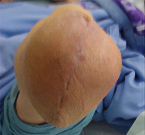
Posterior flap
Skew flap