Thermography and
physiotherapy

What is thermography?

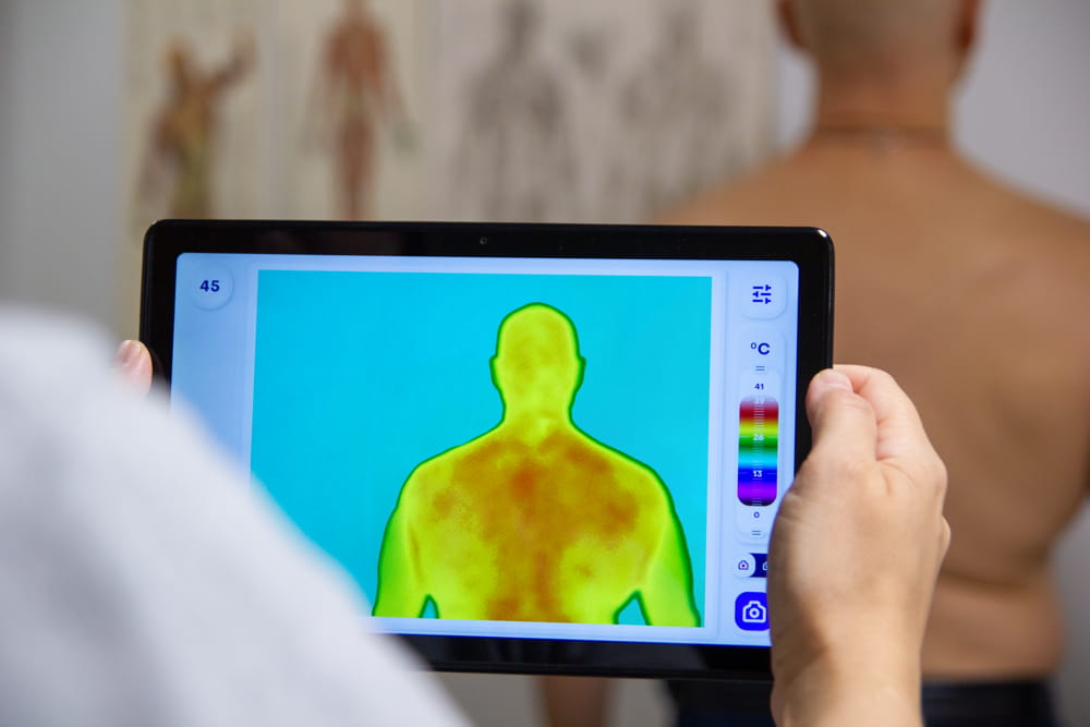
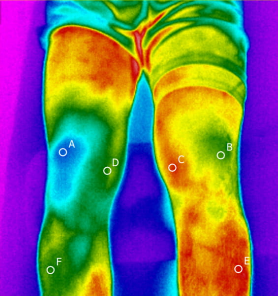
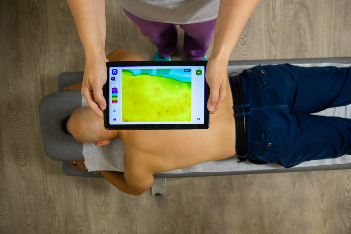
Thermal imaging is a quick and safe method of examining the surface temperature of the skin. Asymmetrical temperature differences or other anomalies indicate abnormalities in the body and are easy to locate using thermal imaging.

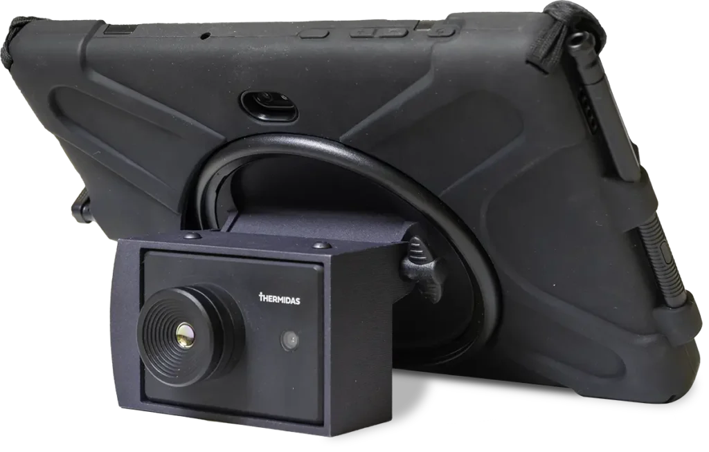
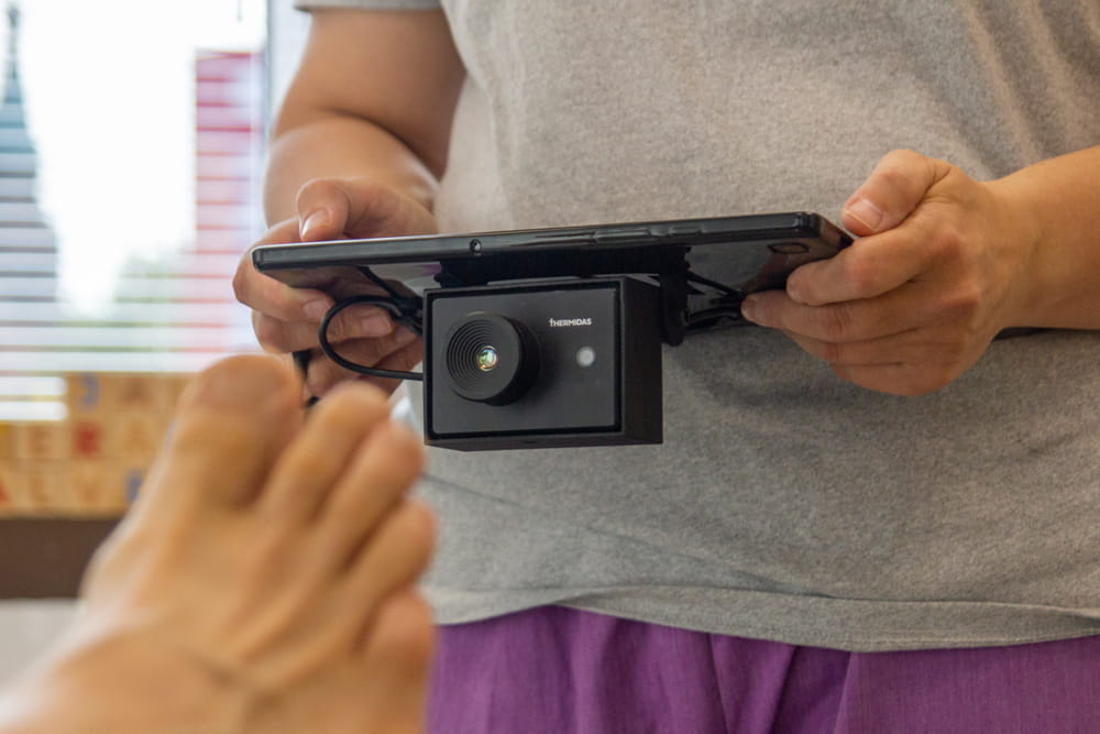
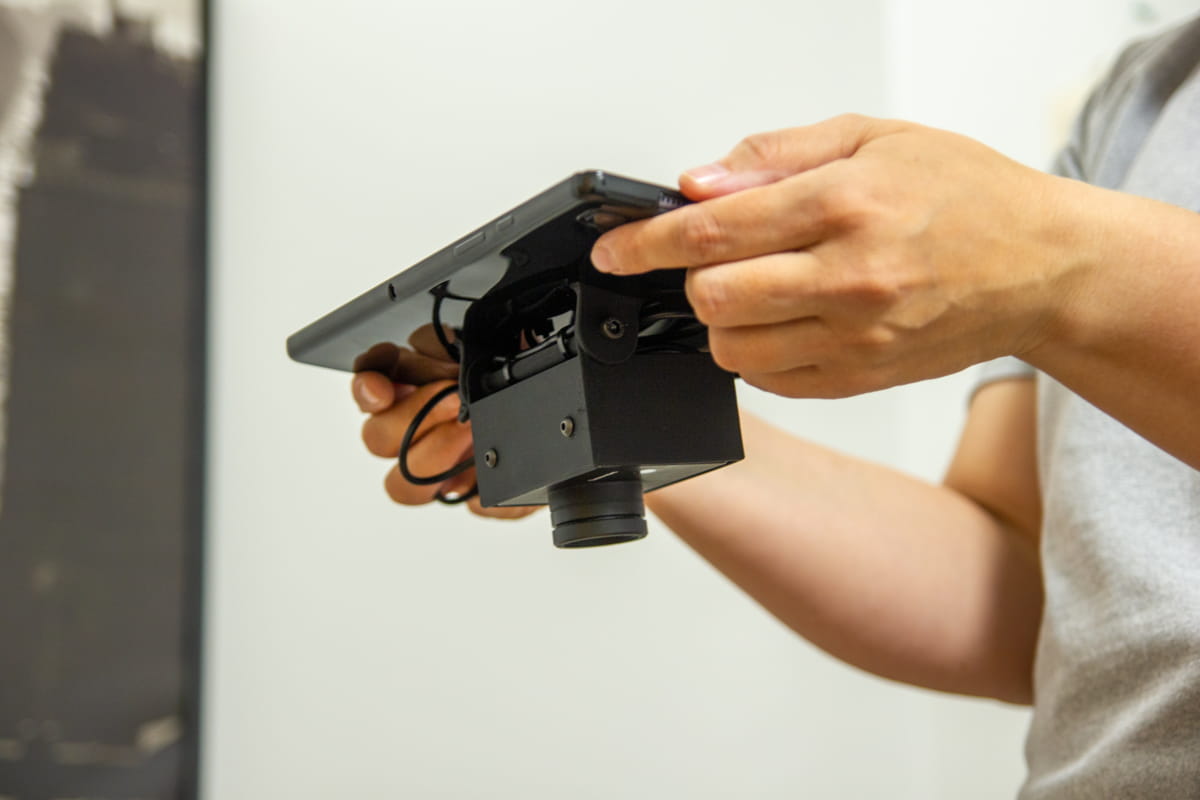
The Thermidas thermal imaging system is a non-invasive, early diagnostic tool to measure the skin’s surface temperature and visualise the results immediately.

Benefits of thermal imaging

Early diagnostics

Assess the location and extent of the problem area. Observe changes before the appearance of clinical symptoms.

Treatment

Monitor the effectiveness of treatment and progress in recovery.

Recovery

Monitor the results of training and the progress of rehabilitation

Example case

Hamstring injury

  • Professional football player was injured during a game
  • Physiotherapist treated with dry needling on the next day
  • Two days after the injury thermal images were taken
  • Right leg is 2 degrees colder and a doctor’s consult was recommended
  • A hamstring injury was diagnosed on the following day

Example case

Fresh ACL injury

  • Top-level football player
  • The patient was in considerable pain
  • The patient could not activate their vastus medialis during a wall sit test
  • The observations support the selection of the correct treatment path and confirm the patient’s story
  • Considerable temperature differences

Recommended solution

IRT-384 Tablet

The IRT-384 is a convenient infrared-enabled tablet that is designed for the daily screening of patients. The high-resolution IR imager quickly highlights temperature differences in skin areas.

Ask for availability.

IRT-384 Tablet in use