Acute Charcot foot has symptoms similar to osteomyelitis, and making the correct diagnosis can be challenging. Blood tests, X-rays or an MRI scan can help with diagnosis, but recent research also offers a quicker and more patient-friendly method to help.
The recently published guidance from the International Working Group on the Diabetes Foot recommends those who are deemed to be at moderate or high risk of foot ulceration (IWGDF risk 2-3) be encouraged to use daily skin temperature monitoring to identify any early signs of foot inflammation and help prevent a first or recurrent plantar foot ulcer (1).
A recent article by Gillian Harkin published in The Diabetic Foot Journal demonstrates the usefulness of thermal imaging as an early diagnostic tool through a patient case study (2).
The paper discusses an interesting case study of a 67-year-old woman with type 1 diabetes who had a previous history of Charcot in both legs. A new ulcer developed on the left leg and started to show signs of inflammation.
On the basis of X-rays and MRI, the patient was started on osteomyelitis treatment and the wound started to heal.
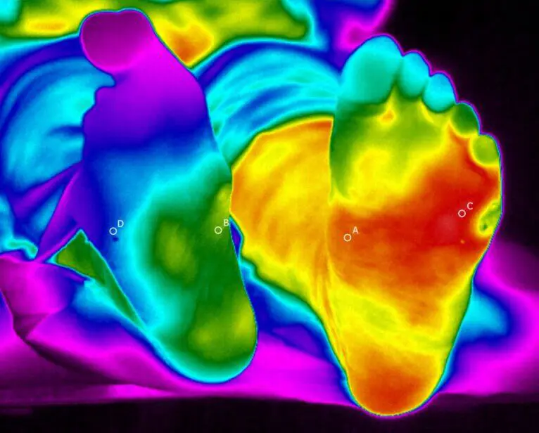
However, new magnetic images taken four weeks later raised suspicions about Charcot reactivation. A temperature difference of 5.4°C observed with a handheld thermometer supported this suspicion and it was decided to immobilise the leg.
The patient’s feet were thermally imaged with a Thermidas ThIR-A615 system. An area of the inflamed leg was found to be 7°C warmer than the healthy one and heat was seen radiating from the wound site. This was not observed in the previous examination, which showed very little evidence of inflammation.
The treatment team agreed that the temperature distribution was consistent with osteomyelitis and not a new case of Charcot. The thermal images also made it easier to explain the problem and justify the treatment to the patient.
After a course of antibiotics and orthosis, the leg temperature stabilised and the wound healed.
Thermal imaging was an essential part of making the right diagnosis and helped to achieve a better treatment outcome.
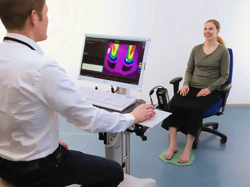
Thermidas ThIR A615
(1) Bus SA, Sacco ICN, Monteiro-Soares M et al (2023) Guidelines on the prevention of foot ulcers in persons with diabetes (IWGDF 2023 update).
(2) Harkin G (2023) Imaging in osteomyelitis and Charcot neuroarthropathy: can infrared thermography aid in diagnosis?. The Diabetic Foot Journal 26(1): 24–8