In recent years, thermal imaging has become more widespread in the health sector and has for example been included in the Charcot foot diagnostic recommendations in Finland.
Tampere University Hospital is one of the pioneers of thermal imaging in Finland. Jorma Lahtela, Senior Physician at TAYS, highlights the importance of thermal imaging in early diagnosis and shares his own experiences of the benefits of the method.
“Thermal imaging is a valuable tool for Charcot foot diagnostics and treatment monitoring, even replacing repeated MRI scans. It can be used, for example, to determine when bone healing has progressed to the point where the use and load on the leg can be increased.”
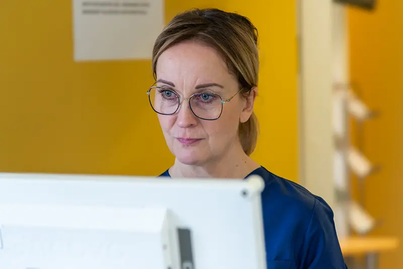
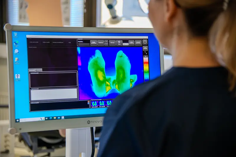
Thermidas’ ThIR-A615 system has also been installed at Kantahäme Central Hospital since 2021. Podiatrist Susanna Satta uses thermal imaging almost daily in her work.
Satta takes thermal images of the feet of Charcot patients every two weeks when the plaster cast is changed. The treatment usually lasts for about a year and regular imaging continues until the end of the treatment.
New uses for the system have been found even after the actual treatment phase. Thermal imaging also makes it easy to assess the fit of orthopedic footwear.
“In many cases, the neuropathy is so bad that the person is unable to recognize the forming of an abrasion.”
The customization of footwear and insoles becomes much easier when the assessment of fit is not left to sensory perception.
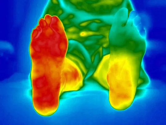
For example, a wound in the heel area can easily cause a person to put more weight on the front of the foot.
“With a thermal imaging system, you can detect things that can’t be seen with the naked eye.”
Early detection allows for faster reaction and appropriate treatment measures to be taken at an early stage.
Before Thermidas’ thermal imaging system, Satta had been using a traditional thermometer for more than twenty years. The differences with the new method have been significant.
“Thermal imaging has totally revolutionized the treatment of the Charcot foot.”
A large proportion of patients are working-age men, and the year-long treatment process can be mentally taxing. Compared to the traditional use of thermometers, thermal imaging has also made a huge difference in terms of treatment adherence.
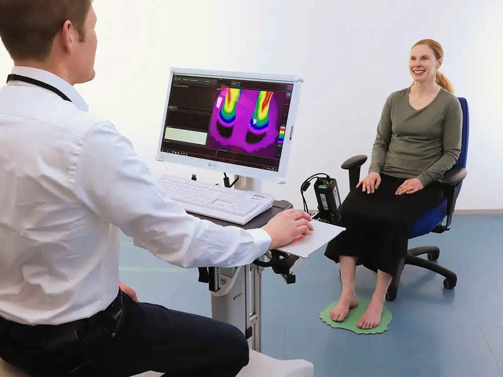
“We can look at thermal images together and compare them with those taken, say, two months ago. The patient can see the progress and understand that the treatment is working. It’s an incredible tool.”
Among other things, several patients who have previously refused plaster treatment have agreed to start treatment after seeing the thermal images.
In two years, the system has become essential to the care of Charcot patients.
“This sort of system should be everywhere where diabetic feet are treated and also in primary care to screen for high-risk feet.”