
Where prevention breaks down
Three reasons a resident does not report the problem
Pressure damage announces itself through discomfort. In residential care, the residents at highest risk are precisely those least able to send that signal - so the first indication arrives when the skin has already broken.
01
Sensation is reduced. Diabetic neuropathy, spinal injury and post-stroke paralysis all remove the pain that would otherwise prompt a resident to shift position.
02
Communication is limited. Advanced dementia, aphasia and non-verbal disability leave a change in behaviour as the only available clue, and behaviour has many causes.
03
Early change is not visible. Pressure and shear over bony prominences happen slowly over time, but become visible only when the damage is already done.
What thermal imaging adds
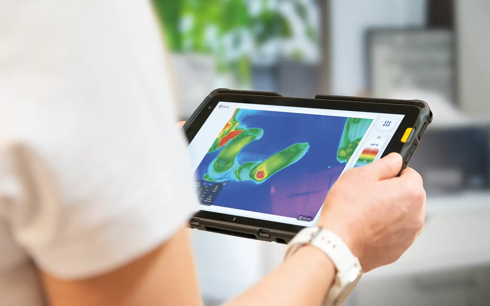
An objective assessment tool. Thermal imaging provides clear physiological data that doesn't rely on visual inspection or risk tools.
More time to react. Thermal patterns associated with inflammation and pressure stress can be detected before clinical symptoms.
The resident's own baseline. Change is read against their earlier images, not against a population average.
Thermal imaging supports assessment and monitoring. It does not diagnose, and findings are always interpreted by a healthcare professional.
Peer-reviewed proof
The evidence behind pressure injury detection
Thermal imaging for pressure injury prevention has been tested in intensive care units, hospital wards and nursing homes, and published in clinical journals. The studies explain and prove the mechanisms, and the Mehiläinen case further down is a real-world example of effective PI prevention.
13×
higher risk of pressure injury on the day before anything is visible to the eye. Measured in 263 hospitalised patients, with near-perfect discrimination (AUC 0.98).
Jiang et al., British Journal of Dermatology, 2022
A day before the skin shows it
In a prospective cohort of 263 hospitalised patients, a thermography-based model detected pressure injuries a full day before they became visually apparent.
More accurate than the scale you already use
In 415 intensive care patients monitored daily for ten days, thermography of the sacral area predicted pressure injury better than the Braden scale.
Independent of skin tone
A 2025 review found hypothermic areas of non-blanching erythema are 31.8× more likely to progress to necrosis, and that thermal assessment works regardless of skin colour — removing a known blind spot in visual checks.
A clear image meant nothing developed
Across 114 consecutive ICU admissions, thermal imaging found 12 anomalies invisible to clinical assessment, and the false-negative rate was zero: none of the 105 patients with a clear thermal baseline went on to develop a visible deep-tissue injury.
Thermal imaging complements skin assessment and existing risk scoring rather than replacing either. The Jiang and Cai cohorts were intensive care and hospital populations, not care homes. Koerner et al. declare author payments from a device manufacturer; it is cited here for its zero-false-negative finding, not its financial figures. For how pressure damage develops and what each stage looks like thermally, see our pressure injuries page.
Revealing hidden information
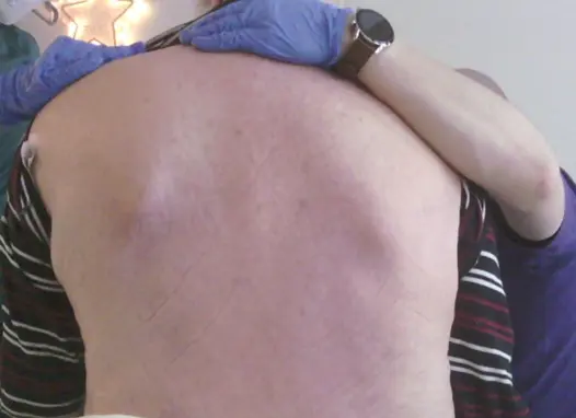
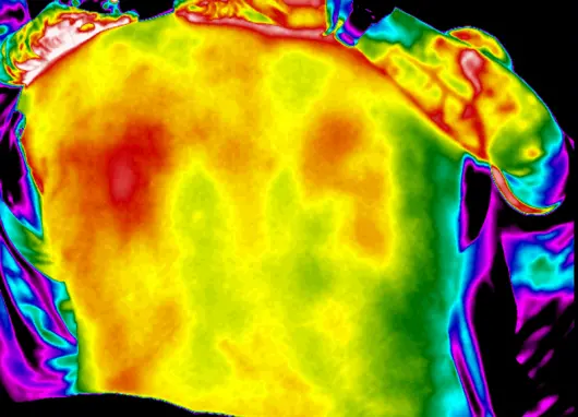
The photograph on the left is what a skin check had to work from
Two cases from two different facilities. In both cases the thermal frame carried information the eye had no access to, and in both cases care changed the same day.
Visual image
Thermal image
Case 01 • Nursing home, Finland
Sensation gone on one side, and no complaint from the resident
A 78-year-old resident, bedridden after a cerebral infarction and paralysed on the left side, imaged during routine physiotherapy rather than in response to any concern.
The thermal image showed a high-risk area on the paralysed side that was not yet visible to the eye.
✔
Assisted sitting and more frequent side changes began that day. No ulcer formed.
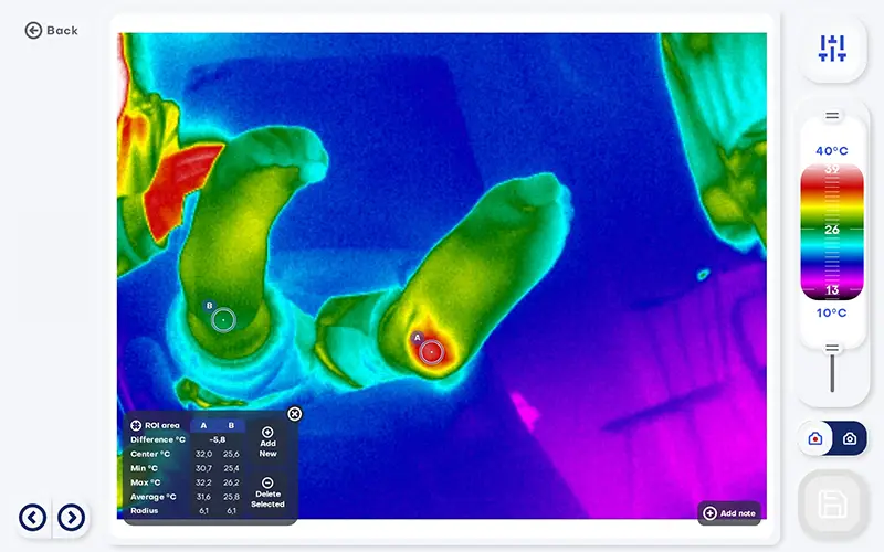
Visual image
Thermal image
Case 02 • Residental care, Germany
A change in behaviour was the only symptom available
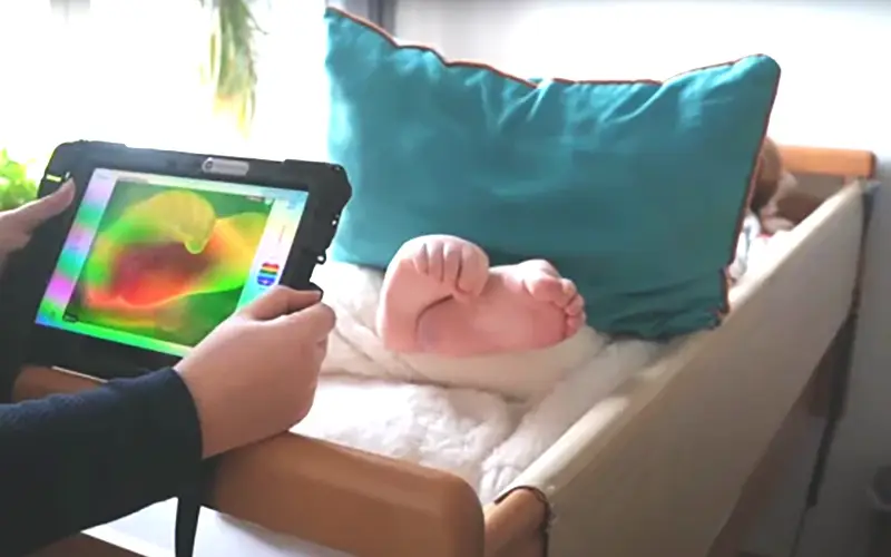
A resident in her mid-thirties with multiple disabilities, non-verbal. Staff noticed she had become less mobile and more distressed, but a skin check found nothing to explain it.
The tablet showed the painful area clearly, turning a behavioural observation into a located finding.
✔
Treatment started in good time, rather than after the skin had broken.
Beyond pressure ulcer prevention
Same method, same device, multiple different applications
Homes that bought it for pressure injuries end up using it for whatever else raises a temperature question.
A wound starting to turn
Raised temperature in the skin around a wound is associated with local infection, often before it looks infected. A prompt to escalate rather than watch and wait.
Whether a wound is actually healing
Residents whose periwound skin was warmer than the wound bed were around five times more likely to heal well. A weekly image gives you a trend rather than an opinion.
Joint pain a resident cannot describe
Thermography detects joint inflammation that clinical examination misses, including in joints without visible swelling or tenderness.
Cold feet and circulation
Side-to-side temperature differences are the most informative signal for circulation problems in the legs and feet. Foot asymmetry is also an established early warning in diabetic foot care.
Published follow-up · Mehiläinen care home, Finland
Over a year with no new pressure ulcers
A four-week pilot in May-June 2024 became part of the daily round that autumn. The home had four to six active pressure ulcers at any given time when the trial started. Six months later the home reported no new ones and at the autumn 2025 review, still none.
Autumn 2024, at adoption
4-6 active
After six months of use
0 new
After one full year
Still 0
Staff reported that thermal imaging is currently the best tool they have for pressure ulcer prevention. It also helps them recognise and manage pain in chronic ulcers and in severely demented residents.
Follow-up report, May 2026 · pilot run with Mehiläinen and the Wellbeing Services County of Pirkanmaa under the DH2 project.
Real-world results
“We were able to make faster medical decisions, resulting in better outcomes, decreasing the use of expensive diagnostic exams, and in one case avoid a toe amputation.”

Fiona Main
NHS Highland
Almost 80 % of Pressure Ulcers are not reported, yet absorb 55 % of costs in the UK
»
The Care Quality Commission requires reporting of Stage 3 and 4 pressure ulcers under statutory duty of candour
»
Some 55 % of care home costs related to pressure ulcers are spent treating Stage 1 and 3 Pressure Ulcers
»
Manual skin checks are prone to human error, often resulting in missed early signs and delayed treatment
»
Early detection is key - advanced-stage ulcers can be prevented by promptly identifying and managing Stage 1 and Stage 2 Pressure Ulcers

Assumes UK Care Home residents = 441,479
Recommended solution
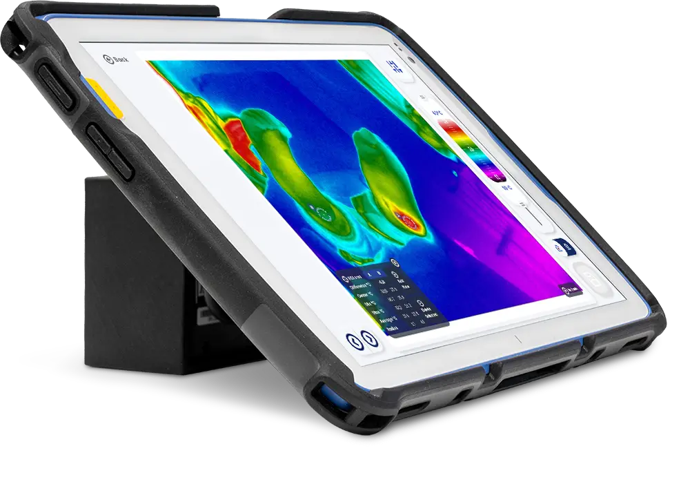
IRT-384 Tablet - the whole workflow in your hands
The IRT-384 Tablet is a portable device for capturing and analyzing thermal images. Works at the bedside, in the clinic room, and during a home visit with the same protocol.
Thermal imaging also supports the client’s understanding of rehabilitation progress by providing clear, objective information on the effects of treatment.
Get started
Take a step toward preventive care
We are here to help you. Let us know whether you have any questions, want to see an online demo first, or prefer to start thermal imaging with a trial.
You can also email or call us:
info@thermidas.fi
+358 20 794 0961
